
Use our bioimaging unit

Explore our approaches to imaging biological and non-biological samples.

An overview of the unit

What we do, the types of projects for which our equipment can be used, and the support you can expect during your research.


Our instruments form a part of a wider imaging network within Bath. Learn more about the Light Microscopy Research Network at Bath.

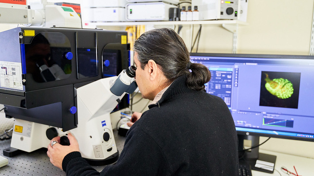
Our unit encompasses several approaches to imaging biological and non-biological samples. The common denominator of these is the use of a light source, usually in the visible spectrum (photons), to produce a signal, which is then read by a suitable detector (our eyes or a specialised electronic device). This is in contrast to electron microscopy, where the 'light' source is actually a beam of electrons.

Our bioimaging instruments are used in a variety of projects, including studies of inflammatory diseases, cancer research, stem cell biology, diabetes, neuroscience, developmental biology, and drug discovery.

Training and assistance are provided at all levels, including guidance on planning your imaging experiments and preparing your samples, and help with image and data analysis.


Light microscopy

Learn about light microscopy and the equipment we can offer.


Light microscopy refers to all the techniques that employ a light source in the visible spectrum and a microscope, to view tiny objects, usually smaller than 1mm. There are currently two major types of light microscopy recognised: Brightfield and Fluorescent microscopy.

Hypoxic facility

Our equipment and how it can support your hypoxic and anoxic research.


With this facility, we can control oxygen levels to study different aspects of cell function, from a single cell to entire cell populations. This provides cell models that mimic oxygen levels in living systems, enabling you to explore the effects of oxygen deprivation, which can lead to tissue damage and disease.

Enquiries

If you have any questions, please contact us.