Use our light microscopy equipment

Find out what light microscopy is and the equipment we can offer.

Our equipment

Light microscopy refers to all the techniques that employ a light source in the visible spectrum and a microscope to view tiny objects. There are currently two major types of light microscopy: Bright-field and Fluorescent microscopy.


We hold these pieces of equipment:

  • Zeiss LSM 880 confocal system with Airyscan and Multi-photon laser
  • Zeiss Z.1 LightSheet
  • Leica Dmi8 widefield system

The confocal laser scanning microscopes enable researchers to create detailed 3D pictures of cell organelles, and to examine ‘live’ cells through incubation systems that facilitate the study of cellular changes over time.


Zeiss LSM 880 confocal system with Airyscan and Multi-photon laser

Based on a Zeiss Axio Observer Z1, the Zeiss LSM 880 is a fully motorised, multi-modal inverted microscope system that you can book as part of your research.


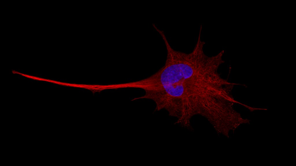
A neuron in orthogonal projection

The system offers single or two-photon excitation, spectral emission detection, multi-photon imaging, and super resolution with an Airyscan detector.

The microscope is equipped with seven excitation laser lines:

  • 405 nanometres
  • 458 nanometres
  • 488 nanometres
  • 514 nanometres
  • 561 nanometres
  • 594 nanometres
  • 633 nanometres

The Zeiss LSM 880 is also equipped with an InSight DS+ Ti:Sapphire femtosecond pulsed laser (tunable between 690 and 1,300 nanometres), being able to excite the vast majority of the commercially available fluorophores.

Depending on the application, emission is detected internally by a 34-channel detector array or the Super Resolution Airyscan detector, and externally by two PMTs at the transmitted path or a BIG.2 detector at the reflected path.

A variety of air, oil, and water objectives offer flexibility in imaging diverse samples. The offered imaging applications cover the biological, physical and material sciences including, but not limited to:

  • multi-colour confocal imaging
  • multi-colour 3D imaging
  • 4D imaging (x, y, z, and time)
  • live cell imaging
  • multi-photon (two-photon) imaging
  • Second/Third Harmonic Generation
  • steady-state Förster/fluorescence resonance energy transfer (FRET)
  • fluorescence recovery after photobleaching (FRAP)
  • spectral unmixing and fingerprinting
  • co-localisation studies
  • calcium imaging or other ratio imaging
Zeiss Z.1 Lightsheet microscope

The Zeiss Z.1 is a fully motorised (X, Y, Z, 360o rotation), multimodal single plane illumination microscope (SPIM) system.


The system is ideal for fast and gentle long-term 3D imaging of large living samples (zebrafish, organoids, small embryos), and for 3D imaging of fixed and/or cleared thick samples up to 1 centimetre (small brains, organs, larger embryos).

The system offers 5 times and 10 times illumination optics and 5 times, 10 times and 20 times detection optics with sample chambers for live samples (RI 1.33) and for cleared samples (RI 1.45). Excitation is achieved through 6 solid state lasers:

  • 405 nanometres
  • 445 nanometres
  • 488 nanometres
  • 514 nanometres
  • 561 nanometres
  • 638 nanometres

The five filterset modules allow imaging of a wide range of fluorescent tags. Illumination can be single-sided or dual-sided (with online Dual-Side fusion for enhanced and illumination of the sample), with the added option of Pivot Scanning to reduce shadowing and striping artifacts.

Image acquisition is achieved by two high-resolution PCO.edge 16bit sCMOS cameras, offering simultaneous two-channel detection.

The system is networked with an analysis workstation equipped with the Arivis Vision4D software for advance 3D/4D rendering applications and analysis routines.


Leica Dmi8 inverted microscope widefield system

The Leica Dmi8 is a fully motorised, multi-modal inverted microscope system.


It offers various transmitted light illumination techniques:

  • Bright-field
  • phase contrast
  • Differential Interference Contrast (DIC)
  • cross polarisers
  • epi-fluorescence

This widefield system is housed within a hypoxic chamber, offering full environmental control and is ideal for live cell imaging. There are filtersets for classic blue, green, red, and far-red stainings, as well as for CFP/YFP combinations (FRET imaging), and calcium imaging with FURA2.

Contact us

If you have any questions, please get in touch.